Article

Large-bore Vascular Closure: New Devices and Techniques

Register or Login to View PDF Permissions
Permissions× For commercial reprint enquiries please contact Springer Healthcare: ReprintsWarehouse@springernature.com.

For permissions and non-commercial reprint enquiries, please visit Copyright.com to start a request.

For author reprints, please email rob.barclay@radcliffe-group.com.
Average (ratings)
No ratings
Your rating

Abstract

Endovascular aneurysm repair, transcatheter aortic valve implantation and percutaneous mechanical circulatory support systems have become valuable alternatives to conventional surgery and even preferred strategies for a wide array of clinical entities. Their adoption in everyday practice is growing. These procedures require large-bore access into the femoral artery. Their use is thus associated with clinically significant vascular bleeding complications. Meticulous access site management is crucial for safe implementation of large-bore technologies and includes accurate puncture technique and reliable percutaneous closure devices. This article reviews different strategies for obtaining femoral access and contemporary percutaneous closure technologies.

Disclosure:The authors have no conflicts of interest to declare.

Received:

Accepted:

Correspondence Details:Nicolas Van Mieghem, Department of Interventional Cardiology, Thoraxcenter, Erasmus MC, Office Nt 645, Dr Molewaterplein 40, 3015 GD Rotterdam, the Netherlands. E: n.vanmieghem@erasmusmc.nl

Copyright Statement:

The copyright in this work belongs to Radcliffe Medical Media. Only articles clearly marked with the CC BY-NC logo are published with the Creative Commons by Attribution Licence. The CC BY-NC option was not available for Radcliffe journals before 1 January 2019. Articles marked ‘Open Access’ but not marked ‘CC BY-NC’ are made freely accessible at the time of publication but are subject to standard copyright law regarding reproduction and distribution. Permission is required for reuse of this content.

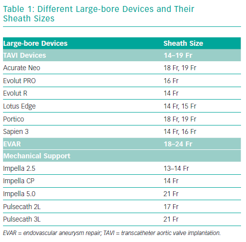
Minimally invasive procedures such as endovascular aneurysm repair (EVAR), transcatheter aortic valve implantation (TAVI) and implantation of mechanical circulatory support (MCS) are gaining ground on traditional surgery.1–3 These procedures require large-bore access, which is inherently associated with vascular complications and bleeding. Despite the reduction in size of these devices (Table 1), vascular- and bleeding complications are frequent and are reported as high as 20% in TAVI and 12–22% in EVAR.4–7 These adverse events lead to prolonged hospitalisation, the need for packed cell transfusion and an increased short and longer-term mortality.8 Common risk factors for access site complications are female sex, extremes of weight, renal insufficiency and anticoagulation use.9–10 This article focuses on strategies for femoral access and closure when using large-bore devices.

Obtaining Access

Good closure starts with good access. The ideal puncture site is located in the common femoral artery between the inferior border of the inferior epigastric artery (IEA) that marks the retroperitoneal space and above the femoral bifurcation. Punctures that are too high are non-compressible and are associated with retroperitoneal bleeding.11 Punctures below the femoral bifurcation, in a small calibre artery, are unsuitable for large-sized sheaths used in EVAR, TAVI and mechanical LV support and should not be closed with percutaneous closure devices per respective instructions for use. There are different strategies for obtaining peripheral access for large-bore devices.

Anatomical Landmarks

When using anatomical landmarks, the operator identifies the inguinal ligament by connecting the anterior-superior iliac spine with the symphysis pubis. Under palpation of the femoral pulse, the needle is inserted into the common femoral artery just below the imaginary line of the inguinal ligament. This strategy is highly dependent on operator experience, which is dropping with increasing numbers of procedures performed via the radial artery – the so-called radial paradox.12,13 A retrospective study by Pitta et al. found that in approximately 13% of the cases, the actual access site was located outside the optimal location, when solely anatomical landmarks were used for puncture guidance. Access outside the target location was associated with more vascular complications.14

Ultrasound-guided Access

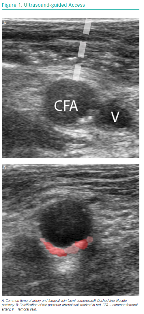
Ultrasound-guided access is performed using a linear ultrasound probe. The first step is to visualise the common femoral artery bifurcation in a longitudinal view to determine the exact bifurcation location and extent of arterial wall calcifications. The probe is then turned counter-clockwise to get a cross-sectional view of the femoral artery above the bifurcation. Vein and artery are distinguished by means of compression. The femoral artery is punctured under a 45° angle, the correct needle pathway and vessel entry is confirmed by ultrasound (Figure 1).

Ultrasound-guided access precludes radiation and is easy to apply after a steep learning curve. It provides a real-time image of the puncture site of interest. Ultrasound allows:

  • the differentiation of non-compressible and pulsatile arteries from compressible veins;
  • the identification of the femoral bifurcation;
  • the appreciation of the degree, location and distribution of calcifications and the selection of a puncture site without anterior wall calcification;
  • the monitoring of needle entry into the vessel, avoiding side or posterior wall puncture.15

Different Large-bore Devices and Their Sheath Sizes

Article image

Compared with fluoroscopic guidance, ultrasound guidance reduces the number of attempts and median time to access.16

Fluoroscopic-guided Access

Fluoroscopic-guided access assumes a consistent spatial relationship between the common femoral artery and femoral head.17 Under X-ray, a radiopaque instrument, such as a haemostat or puncture needle, is placed over the femoral head to locate the appropriate height for puncture. Assumptions may be inaccurate in patients with high femoral bifurcations. Alternatively, a wire or (e.g. pigtail) catheter can be inserted from a contralateral access and navigated towards the level of the ipsilateral femoral head to serve as a target for the fluoroscopy-guided puncture. A small contrast injection through the pigtail catheter may further map the common femoral artery and serve as a bull’s eye for the operator. Fluoroscopy-guided arterial puncture is effective and associated with a low incidence of vascular complications, but it has not been shown to be superior to the use of anatomical landmarks.18–20 The major downside of this technique is its reliance on radiation, in particular to the operator’s hands.

Surgical Cut-down

Surgical cut-down can expose the common femoral artery and allows for direct-vision access and allows for direct-suture closure. Surgical cut-down is associated with a longer procedure time, increased length of hospitalisation and more wound infections.21–23 Complications seem to occur less frequently when an oblique incision is chosen over a vertical incision.24

Vascular Closure

Surgical Closure

In principle, a surgical suture technique is applied for closure after surgical cut-down for femoral access. Surgical cut-down and closure increases the chance for wound infection or iatrogeneous femoral nerve damage. At present, most TAVI and EVAR procedures are performed in a total percutaneous matter, but a surgical cut-down may still be preferred in selected patients, such as the very obese or those with femoral grafts or stents.25

Ultrasound-guided Access

Article image

Suture-based Closure Devices

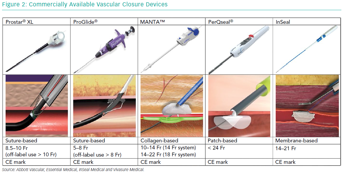
The vast majority of large-bore vessel closure is performed by percutaneous suture- based techniques like the Prostar® XL and multiple ProGlide® (Abbott Vascular) vascular closure devices (VCD) (Figure 2). Both devices are predominantly inserted using a pre-closure technique.

Commercially Available Vascular Closure Devices

Article image

Forest Plot Showing Odds Ratio for Any Bleeding and Any Vascular Complication

Article image

The Prostar XL device is inserted over a guidewire. Its position in the artery is confirmed when pulsatile flow evades the main tube of the device. Four pre-prepared sutures inside the device are pulled out while maintaining the device in the same position. This allows four needles to be pulled back, leaving the sutures in place. The arteriotomy can be closed by pulling the sutures and closing the knots.

The ProGlide technique typically requires two devices for large-bore arteriotomies. The devices are inserted before the procedure and are deployed at the 10 o’clock and 2 o’clock position. After the procedure is concluded, the introducer sheath is removed. The sutures are approximated and the vessel wall is closed.26 Both suture-based techniques can be executed with a safety wire in place in order to use additional suture- or plug-based closure devices if there is incomplete arteriotomy closure.

ProGlide was originally introduced to clinical practice for small-bore arteriotomy closure, but its use was extended to EVAR, first under surgical cut-down and later in a completely percutaneous fashion.27 Compared to surgical cut-down, there are fewer groin complications when using a VCD, and the procedural time is shorter (91 minutes ± 32 versus 153 minutes ± 112; p<0.05).28,29

A propensity matched analysis in TAVI patients by Barbash et al. showed lower rates of major vascular complications with use of ProGlide compared to Prostar XL (1.9% versus 7.4%; p<0.001) and lower rates of major (3.2% versus 16.7%; p<0.001) and minor bleedings (8.9% versus 13.6%; p=0.032).7 Conversely, a study in an Italian hospital reported more vascular complications with ProGlide versus Prostar XL closure (24.0% versus 11.4%; p=0.007).30 Basically, local experience will determine suture-based closure success and it is recommended that each operator or centre adopts and masters one suture-based technique.

Suture-based closure has also been successfully applied for closure of axillary and subclavian arteriotomies.31,32

Collagen-based Closure

The MANTA VCD (Essential Medical) is a collagen-based closure device (Figure 2). It consists of a poly-lactic coglycolic toggle within the artery, connected to a bovine collagen plug, exterior to the vessel wall. A stainless-steel lock is tampered down pushing the collagen and toggle together in order to sandwich the arterial puncture site between the toggle and the collagen. The proper amount of tension that the operator has to apply is indicated by the appearance of a green marker on the device handle. The toggle and collagen plug resolve completely in 6 months.33 The MANTA has a 14 Fr and 18 Fr version for arteriotomy closure between 10 and 14 Fr and 14 and 22 Fr, respectively, and obtained the CE mark in 2016. The MANTA has also been applied for completely percutaneous closure of axillary arteriotomies after TAVI.34,35

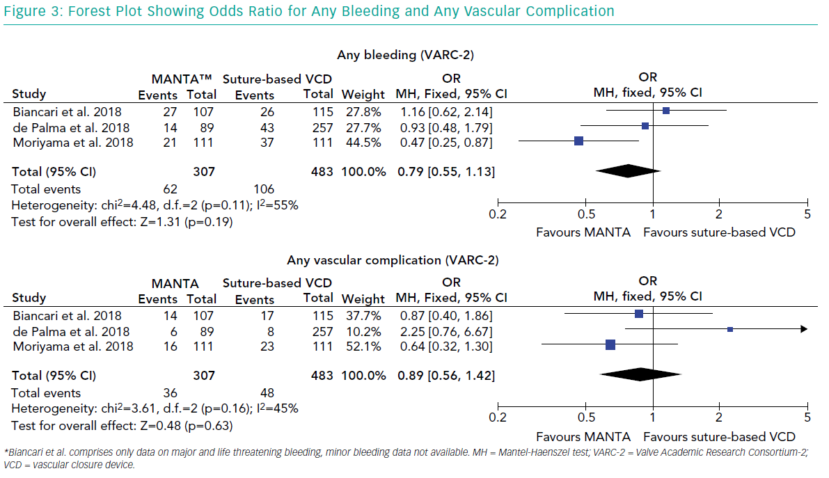
There are no randomised head-to-head comparisons, but retrospective data show lower bleeding complications and comparable vascular complications with the MANTA device compared to the Prostar XL (Major bleeding 2.3% versus 9.3%; p=0.03; major vascular 2.3% versus 0.4%; p=0.48).36 A propensity matched analysis by Moriyama et al. confirmed less VARC-2 bleeding (18% versus 33%; p=0.01) but no difference in vascular complications (14% versus 21%; p=0.21) with MANTA.37 Biancari et al. found no significant difference between the MANTA and ProGlide in terms of bleeding (22% versus 25%; p=0.469) or major vascular complications (12% versus 9%; p=0.498)38 (Figure 3).

The MANTA device has a short mean time to haemostasis, ranging from 22 seconds to 2 minutes 23 seconds. There is no comparable time to haemostasis data for other VCDs.35,36,39

Miscellaneous

Other novel dedicated large-bore closure devices include the InSeal (InSeal Medical) and PerQseal® (Vivasure Medical) VCD (Table 2).

The InSeal VCD is a membrane-based device consisting of a self-expanding nitinol frame, a biodegradable membrane and a bioresorbable polyglycolic acid (PGA) tether. The InSeal device is introduced with the membrane in a collapsed configuration. The sheath is then pulled back and the release wire is pulled to deploy the VCD. The membrane is pushed against the arteriotomy site by the nitinol frame and traction is kept by keeping the tether fixed to the skin using a steristrip or suture. The flexible membrane should compensate for arterial wall irregularities and calcifications. The specially designed frame allows re-access within 26 weeks. The first in human experience showed technical and therapeutic success in all nine cases.40 Unpublished post CE mark clinical experience showed a mean time to haemostasis <1 minute and 0% Major VARC-2 vascular complications and 7.7% bleeding complications in a series of 52 patients.41

The PerQseal VCD consists of a flexible intravascular patch supported by a scaffold. The surface of the patch is textured to promote adherence to the vessel wall. An external locator extends through the arteriotomy, which keeps the patch in place. The implant is fully absorbable after 180 days. It received the CE mark in 2016. In 120 patients, from the unpublished Frontier series of studies, including TAVI, EVAR and thoracic endovascular aortic repair, no major vascular complications occurred.42

Conclusion

The use of large-bore arteriotomies is peaking with the expanding market of structural heart interventions and MCS. Indeed, catheter-based techniques, such as EVAR and TAVI have greatly replaced conventional surgical operations. Optimal access site management, including the proper puncture and arteriotomy closure technique, is pivotal to secure procedural safety and will ultimately determine the success of catheter-based therapies in clinical practice.

References

  1. Greenhalgh RM, Brown LC, Kwong GP, et al. Comparison of endovascular aneurysm repair with open repair in patients with abdominal aortic aneurysm (EVAR Trial 1), 30-day operative mortality results: randomised controlled trial. Lancet. 2004;364:843–8.
    Crossref | PubMed
  2. Prinssen M, Verhoeven EL, Buth J, et al. A randomized trial comparing conventional and endovascular repair of abdominal aortic aneurysms. N Engl J Med. 2004;351:1607–18.
    Crossref | PubMed
  3. Durko AP, Osnabrugge RL, Van Mieghem NM, et al. Annual number of candidates for transcatheter aortic valve implantation per country: current estimates and future projections. Eur Heart J. 2018;39:2635–42.
    Crossref | PubMed
  4. Carroll JD, Vemulapalli S, Dai D, et al. Procedural experience for transcatheter aortic valve replacement and relation to outcomes: the STS/ACC TVT Registry. J Am Coll Cardiol. 2017;70:29–41.
    Crossref | PubMed
  5. Linke A, Wenaweser P, Gerckens U, et al. Treatment of aortic stenosis with a self-expanding transcatheter valve: the International Multi-centre ADVANCE Study. Eur Heart J. 2014;35:2672–84.
    Crossref | PubMed
  6. Nelson PR, Kracjer Z, Kansal N, et al. A multicenter, randomized, controlled trial of totally percutaneous access versus open femoral exposure for endovascular aortic aneurysm repair (the PEVAR Trial). J Vasc Surg. 2014;59: 1181–93.
    Crossref | PubMed
  7. Barbash IM, Barbanti M, Webb J, et al. Comparison of vascular closure devices for access site closure after transfemoral aortic valve implantation. Eur Heart J. 2015;36:3370–9.
    Crossref | PubMed
  8. Doyle BJ, Ting HH, Bell MR, et al. Major femoral bleeding complications after percutaneous coronary intervention: incidence, predictors, and impact on long-term survival among 17,901 patients treated at the Mayo Clinic from 1994 to 2005. JACC Cardiovasc Interv. 2008;1:202–9.
    Crossref | PubMed
  9. Sherev DA, Shaw RE, Brent BN. Angiographic predictors of femoral access site complications: implication for planned percutaneous coronary intervention. Catheter Cardiovasc Interv. 2005;65:196–202.
    Crossref | PubMed
  10. Tavris DR, Gallauresi BA, Lin B, et al. Risk of local adverse events following cardiac catheterization by hemostasis device use and gender. J Invasive Cardiol. 2004;16:459–64.
    PubMed
  11. Illescas FF, Baker ME, McCann R, et al. CT evaluation of retroperitoneal hemorrhage associated with femoral arteriography. AJR Am J Roentgenol. 1986;146:1289–92.
    Crossref | PubMed
  12. Rafie IM, Uddin MM, Ossei-Gerning N, et al. Patients undergoing PCI from the femoral route by default radial operators are at high risk of vascular access-site complications. EuroIntervention. 2014;9:1189–94.
    Crossref | PubMed
  13. Azzalini L, Tosin K, Chabot-Blanchet M, et al. The benefits conferred by radial access for cardiac catheterization are offset by a paradoxical increase in the rate of vascular access site complications with femoral access: the campeau radial paradox. JACC Cardiovasc Interv. 2015;8: 1854–64.
    Crossref | PubMed
  14. Pitta SR, Prasad A, Kumar G, et al. Location of femoral artery access and correlation with vascular complications. Catheter Cardiovasc Interv. 2011;78:294–9.
    Crossref | PubMed
  15. Sardar MR, Goldsweig AM, Abbott JD, et al. Vascular complications associated with transcatheter aortic valve replacement. Vasc Med. 2017;22:234–44.
    Crossref | PubMed
  16. Seto AH, Abu-Fadel MS, Sparling JM, et al. Real-time ultrasound guidance facilitates femoral arterial access and reduces vascular complications: FAUST (Femoral Arterial Access With Ultrasound Trial). JACC Cardiovasc Interv. 2010;3:751–8.
    Crossref | PubMed
  17. Dotter CT, Rosch J, Robinson M. Fluoroscopic guidance in femoral artery puncture. Radiology. 1978;127:266–7.
    Crossref | PubMed
  18. Abu-Fadel MS, Sparling JM, Zacharias SJ, et al. Fluoroscopy vs. traditional guided femoral arterial access and the use of closure devices: a randomized controlled trial. Catheter Cardiovasc Interv. 2009;74:533–9.
    Crossref | PubMed
  19. Fairley SL, Lucking AJ, McEntegart M, et al. Routine use of fluoroscopic-guided femoral arterial puncture to minimise vascular complication rates in CTO intervention: multi-centre UK experience. Heart Lung Circ. 2016;25:1203–9.
    Crossref | PubMed
  20. Chinikar M, Ahmadi A, Heidarzadeh A, Sadeghipour P. Imaging or trusting on surface anatomy? A comparison between fluoroscopic guidance and anatomic landmarks for femoral artery access in diagnostic cardiac catheterization. A randomized control trial. Cardiovasc Interv Ther. 2014;29:18–23.
    Crossref | PubMed
  21. Nakamura M, Chakravarty T, Jilaihawi H, et al. Complete percutaneous approach for arterial access in transfemoral transcatheter aortic valve replacement: a comparison with surgical cut-down and closure. Catheter Cardiovasc Interv. 2014;84:293–300.
    Crossref | PubMed
  22. Kadakia MB, Herrmann HC, Desai ND, et al. Factors associated with vascular complications in patients undergoing balloon-expandable transfemoral transcatheter aortic valve replacement via open versus percutaneous approaches. Circ Cardiovasc Interv. 2014;7:570–6.
    Crossref | PubMed
  23. Buck DB, Karthaus EG, Soden PA, et al. Percutaneous versus femoral cutdown access for endovascular aneurysm repair. J Vasc Surg. 2015;62:16–21.
    Crossref | PubMed
  24. Slappy AL, Hakaim AG, Oldenburg WA, et al. Femoral incision morbidity following endovascular aortic aneurysm repair. Vasc Endovascular Surg. 2003;37:105–9.
    Crossref | PubMed
  25. Toggweiler S, Webb JG. Challenges in transcatheter aortic valve implantation. Swiss Med Wkly. 2012;142:w13735.
    Crossref | PubMed
  26. Toggweiler S, Leipsic J, Binder RK, et al. Management of vascular access in transcatheter aortic valve replacement: part 1: basic anatomy, imaging, sheaths, wires, and access routes. JACC Cardiovasc Interv. 2013;6:643–53.
    Crossref | PubMed
  27. Krajcer Z, Howell M. A novel technique using the percutaneous vascular surgery device to close the 22 french femoral artery entry site used for percutaneous abdominal aortic aneurysm exclusion. Catheter Cardiovasc Interv. 2000;50:356–60.
    PubMed
  28. Jahnke T, Schafer JP, Charalambous N, et al. Total percutaneous endovascular aneurysm repair with the dual 6-F Perclose-AT preclosing technique: a case-control study. J Vasc Interv Radiol. 2009;20:1292–8.
    Crossref | PubMed
  29. Lee WA, Brown MP, Nelson PR, Huber TS. Total percutaneous access for endovascular aortic aneurysm repair (“Preclose” technique). J Vasc Surg. 2007;45:1095–101.
    Crossref | PubMed
  30. Barbanti M, Capranzano P, Ohno Y, et al. Comparison of suture-based vascular closure devices in transfemoral transcatheter aortic valve implantation. EuroIntervention. 2015;11:690–7.
    Crossref | PubMed
  31. Schafer U, Ho Y, Frerker C, et al. Direct percutaneous access technique for transaxillary transcatheter aortic valve implantation: “the Hamburg Sankt Georg approach”. JACC Cardiovasc Interv. 2012;5:477–86.
    Crossref | PubMed
  32. van Mieghem NM, Luthen C, Oei F, et al. Completely percutaneous transcatheter aortic valve implantation through transaxillary route: an evolving concept. EuroIntervention. 2012;7:1340–2.
    Crossref | PubMed
  33. van Gils L, Daemen J, Walters G, et al. MANTA, a novel plug-based vascular closure device for large Bore arteriotomies: technical report. EuroIntervention. 2016;12:896–900.
    Crossref | PubMed
  34. De Palma R, Ruck A, Settergren M, Saleh N. Percutaneous axillary arteriotomy closure during transcatheter aortic valve replacement using the MANTA device. Catheter Cardiovasc Interv. 2018;92:998–1001.
    Crossref | PubMed
  35. Van Mieghem NM, Latib A, van der Heyden J, et al. Percutaneous plug-based arteriotomy closure device for large-bore access: a multicenter prospective study. JACC Cardiovasc Interv. 2017;10:613–9.
    Crossref | PubMed
  36. De Palma R, Settergren M, Ruck A, et al. Impact of percutaneous femoral arteriotomy closure using the MANTATM device on vascular and bleeding complications after transcatheter aortic valve replacement. Catheter Cardiovasc Interv. 2018;92:954–61.
    Crossref | PubMed
  37. Moriyama N, Lindstrom L, Laine M. Propensity-matched comparison of vascular closure devices after transcatheter aortic valve replacement using MANTA versus ProGlide. EuroIntervention. 2018; epub ahead of press.
    Crossref | PubMed
  38. Biancari F, Romppanen H, Savontaus M, et al. MANTA versus ProGlide vascular closure devices in transfemoral transcatheter aortic valve implantation. Int J Cardiol. 2018;263:29–31.
    Crossref | PubMed
  39. van Gils L, De Jaegere PP, Roubin G, Van Mieghem NM. The MANTA vascular closure device: a novel device for large-bore vessel closure. JACC Cardiovasc Interv 2016;9: 1195–6.
    Crossref | PubMed
  40. Kambara AM, Bastos Metzger P, Ribamar Costa J et al. First-in-man assessment of the InSeal VCD, a novel closure device for large puncture accesses. EuroIntervention. 2015;10:1391–5.
    Crossref | PubMed
  41. Kornowski R. Large-hole trans-femoral closure – the InSeal device. Presented at TVT 2017, Chicago, 14–17 June 2017.
  42. Popma J. New fully absorbable patch-based large-hole vascular closure device. Presented at TCT 2017, Denver, 29 October–2 November 2017.