Article

Lesion Preparation with Orbital Atherectomy

Register or Login to View PDF Permissions
Permissions× For commercial reprint enquiries please contact Springer Healthcare: ReprintsWarehouse@springernature.com.

For permissions and non-commercial reprint enquiries, please visit Copyright.com to start a request.

For author reprints, please email rob.barclay@radcliffe-group.com.
Average (ratings)
No ratings
Your rating

Abstract

Despite significant improvements in stent design, severe coronary calcification continues to impede adequate stent expansion and is associated with worse clinical outcomes. Angiography is limited in its ability to detect and comprehensively characterise calcified plaque. Intravascular imaging provides information on lesion morphology guiding appropriate treatment strategies. Orbital atherectomy allows for lesion preparation of severely calcified plaque prior to stent implantation. Utilising a unique mechanism of action incorporating centrifugal forces, a standard 1.25 mm eccentrically mounted and diamond-coated burr orbits bi-directionally to ablate calcified plaque. Lesion preparation with orbital atherectomy allows for modification of calcified plaque to facilitate stent expansion.

Disclosure:AJ has received institutional funding (unrestricted education grant) from Philips and Abbott Vascular and has served as a consultant for Philips, Abbott Vascular, Opsens Medical, and Boston Scientific. ZAA received institutional research grants to Columbia University from Abbott and Cardiovascular Systems and is a consultant to Abbott, ACIST Medical Systems, Boston Scientific, Cardinal Health, Medtronic and Canon US. All other authors have no conflicts of interest to declare.

Received:

Accepted:

Correspondence Details:Ziad A Ali, 1700 Broadway, 8th Floor, New York, NY 10019, US. E: zaa2112@columbia.edu

Copyright Statement:

The copyright in this work belongs to Radcliffe Medical Media. Only articles clearly marked with the CC BY-NC logo are published with the Creative Commons by Attribution Licence. The CC BY-NC option was not available for Radcliffe journals before 1 January 2019. Articles marked ‘Open Access’ but not marked ‘CC BY-NC’ are made freely accessible at the time of publication but are subject to standard copyright law regarding reproduction and distribution. Permission is required for reuse of this content.

Coronary calcification has posed challenges to successful revascularisation since the development of percutaneous coronary intervention (PCI).1 Coronary artery calcification worsens clinical outcomes irrespective of the revascularisation strategy.2–4 Despite significant improvements in stent design over the past decades, including an improved ability to deliver a stent, severe calcification continues to impede adequate stent expansion with contemporary PCI. Severe calcification is present in nearly one-third of patients undergoing PCI.5 The degree of coronary lesion calcification impacts stent expansion, which is one of the strongest predictors for in-stent restenosis and stent thrombosis.6–10

Historically, routine use of atherectomy has been reserved for undilatable and uncrossable calcified lesions.11,12 The concept of routine lesion preparation for all heavily calcified lesions prior to stent implantation to prevent stent underexpansion represents a recent change in practice, but is supported by data from intravascular imaging-based studies.6,12–18

Assessment of Coronary Artery Calcification

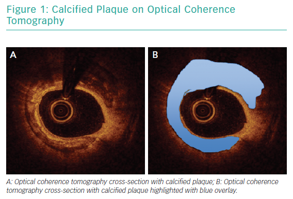
Angiography has been consistently shown to be inadequate for comprehensive assessment of coronary artery calcification.19 In a pivotal analysis of 1,155 coronary lesions, Mintz et al. demonstrated that, while angiography detected calcium in 38% of lesions, intravascular ultrasound (IVUS) was able to detect lesion calcification in 73% of lesions.20 Similar findings were reported in a study that included optical coherence tomography (OCT) in addition to angiography and IVUS.21 Intravascular imaging provides data beyond simple detection of calcified plaque. Lesion morphology, arc of calcification, calcium thickness and length are best assessed with intravascular imaging and can be determinant of the optimal revascularisation strategy.22 Figure 1 illustrates calcified plaque identified on OCT.

Role of Calcium Modification

Mild or moderate calcification is often adequately treated with balloon angioplasty with either a noncompliant balloon, cutting balloon or scoring balloon. When superficial coronary artery calcification is <0.24 mm thick, a balloon alone is often adequate to create fractures in the calcium plate.23 As calcium thickness increases, achieving calcium fracture with balloon angioplasty alone becomes less likely. In patients with an OCT-based calcium score of 4, indicating the presence of calcium thickness >0.5 mm, an arc of calcium exceeding 180o and a continuous length of calcium >5 mm, adjunctive therapies should be used prior to stent implantation.24 Calcium fracture can facilitate improved stent expansion.16,25

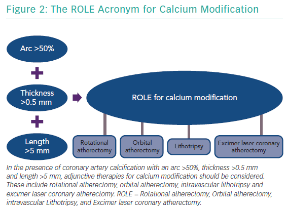
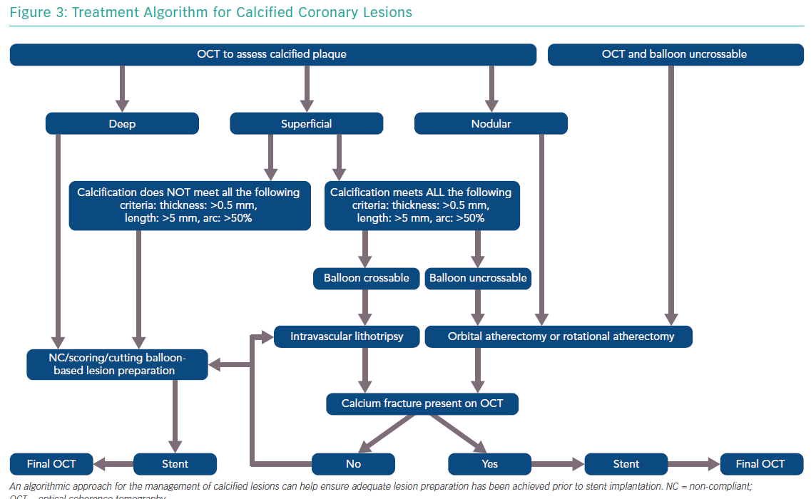
The features of a calcified lesion determine the optimal lesion preparation strategy. The adjunctive therapies that can be utilised for calcium modification as part of PCI can be remembered by the acronym ROLE (Figure 2): Rotational atherectomy, Orbital atherectomy, intravascular Lithotripsy, and Excimer laser coronary atherectomy (ELCA). If a lesion is balloon crossable, intravascular lithotripsy can be used. For lesions that are not balloon crossable, rotational atherectomy or orbital atherectomy are both reasonable options. ELCA can be considered for de novo calcified lesions not amenable to rotational atherectomy, orbital atherectomy or intravascular lithotripsy.26–29 However, the primary role for ELCA in treating calcified plaque is for in-stent restenosis due to an underexpanded stent with significant peri-stent calcification.30,31 An algorithmic approach for the management of calcified lesions can help ensure adequate lesion preparation has been achieved prior to stent implantation (Figure 3). We herein focus on the treatment of calcified plaque with orbital atherectomy.

Calcified Plaque on Optical Coherence Tomography

Article image

The ROLE Acronym for Calcium Modification

Article image

Orbital Atherectomy

Device Setup and Use

Orbital atherectomy for coronary arteries utilises the Diamondback 360® orbital atherectomy system (Cardiovascular Systems) which incorporates a 1.25 mm diamond-coated eccentrically mounted crown that is advanced over a dedicated 0.012" ViperWire® Advance Coronary Guide Wire (Cardiovascular Systems) with a 0.014" tip. ViperSlide® (Cardiovascular Systems) lubricant is infused during treatment to reduce friction from a specialised pump that is mounted on an intravenous pole. The crown is controlled by a hand-held electric powered single-use handle that has two speeds for treatment: low (80,000 rpm) and high (120,000 rpm). The orbital atherectomy system is compatible with a 6 Fr or larger catheter.

Employing centrifugal forces, the 1.25 mm crown orbits as it advances through a coronary artery, allowing for the continuous flow of blood during atheroablation. This unique mechanism of action combined with an average particle size of debris of 2.04 µm – smaller than a red blood cell – may contribute to low rates of no-reflow and transient heartblock with orbital atherectomy.32 A distinguishing feature of orbital atherectomy is its ability to ablate bi-directionally, potentially decreasing the likelihood for burr entrapment.33

The 1.25 mm crown size is standard for all coronary arteries and can treat larger-sized lumens by either increasing the speed (high speed) or by slowing the rate of advancement to allow an increase in the diameter of the elliptical orbit. By decreasing the traverse speed to 1 mm/second at high speed, the burr orbit can exceed 2 mm in diameter.34 Low speed should be used for all initial passes with orbital atherectomy. Following treatment at low speed, if an adequate endpoint has not been reached, treatment can be escalated to high speed. High speed is not necessary for all cases, and specifically should be avoided in vessels <3 mm in diameter or excessively tortuous vessels to reduce the risk for perforation. The GlideAssist® feature allows for rotation at 5,000 rpm and may be used to aid in the delivery and removal of the device but should not be used for treating a lesion. GlideAssist can facilitate the use of orbital atherectomy by a single operator.35

The goal of lesion preparation is to modify calcified plaque, changing the vessel compliance to facilitate stent expansion.36 The slower the burr is advanced with orbital atherectomy, the larger the orbit is and greater the likelihood of achieving plaque modification as it is a time dependent therapy. The burr should be advanced at 1–3 mm/second. Notably, optimal orbital atherectomy technique differs from the standard rapid pecking motion that is characteristic with rotational atherectomy use.

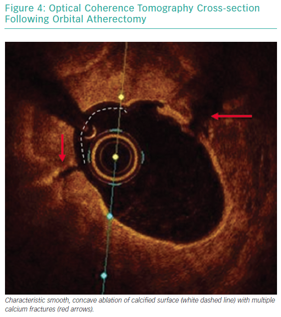
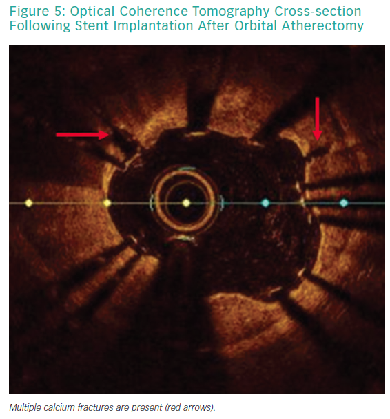
Each pass with orbital atherectomy should be limited to less than 30 seconds. After 25 seconds of continuous treatment, the system emits an audible alert and the crown should be moved to a location outside of the lesion. Rest periods between treatment runs should be for a minimum of duration equal to the preceding run. If the patient becomes symptomatic during treatment, greater rest periods should follow. During atheroablation, pitch changes can be appreciated as the crown engages the hard, calcified plaque. As treatment continues, the pitch change becomes reduced, and tactile resistance decreases.33 Treatment with orbital atherectomy should continue until audible and tactile changes are appreciated, indicating the optimal endpoint has been reached. There is no predefined number of treatment runs that is ideal, and therapy should be adjusted as needed for each specific lesion. While patients have had favourable outcomes following direct stenting after orbital atherectomy, direct stenting in calcified lesions should not be used unless intravascular imaging is performed following lesion preparation confirming an adequate endpoint has been achieved with significant calcium fracture.37 Calcium fracture and the typical smooth, concave ablation seen with orbital atherectomy is readily identified with OCT imaging (Figures 4 and 5).

Clinical Data

The first in-human study of coronary orbital atherectomy of 50 patients treated at two centres in India in 2008 was the Safety and Feasibility of Orbital Atherectomy for the Treatment of Calcified Coronary Lesions (ORBIT I) study, which demonstrated device success of 98% with procedural success in 94% of patients.38 Long-term follow-up was reported and the cumulative major adverse cardiac event (MACE) rate at 3-years follow-up was 18.2%.39 Of note, the ORBIT I investigational study occurred prior to device approval and evaluated four different sized burrs, with only 18.4% of patients having been treated with the 1.25 mm burr that is currently available.38

The pivotal Evaluate the Safety and Efficacy of OAS [Orbital Atherectomy System] in Treating Severely Calcified Coronary Lesions (ORBIT II) study evaluated the impact of orbital atherectomy in 443 patients with severe coronary artery calcification treated at 49 sites in the US from 2010 to 2012.40 The primary safety endpoint, freedom from 30-day MACE, was met in 89.6% of patients and led to device approval in the US in 2013.40 At 3-year follow-up, the overall cumulative MACE rate was 23.5%, and was comprised of 6.7% cardiac death, 11.2% MI and target vessel revascularisation (TVR) in 10.2% of patients with severe calcification treated with orbital atherectomy.41

Treatment Algorithm for Calcified Coronary Lesions

Article image

A real-world multicentre all-comer analysis reported outcomes of 458 patients treated with orbital atherectomy from 2013 to 2015.42 This analysis included a complex cohort of patients that had clinical features that were excluded from the ORBIT II study including unprotected left main disease, low ejection fraction, chronic kidney disease and acute coronary syndrome. Angiographic complications were low with dissection, perforation and no-reflow occurring in less than 1% of cases.42 At 1-year follow-up, the composite rate of major adverse cardiac and cerebrovascular events occurred in 12.6%, with 4.0% all-cause mortality, 1.8% MI rate and TVR rate of 7.5%.43

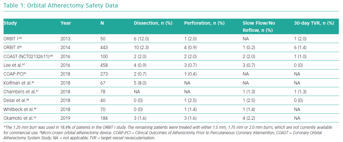
Multiple studies have reported safety data related to orbital atherectomy in both stand-alone orbital atherectomy studies and in observational studies with comparison to rotational atherectomy. The rates of dissection, perforation, slow/no-reflow and 30-day TVR in patients treated with orbital atherectomy are presented in Table 1.

Imaging-based Insights

In a small, multi-centre OCT-based mechanistic study comparing orbital atherectomy with rotational atherectomy in 60 patients, the investigators found that orbital atherectomy creates greater calcium modification in lesions with larger lumen area.51 Calcium fracture following stent implantation occurred in the majority of cases regardless of atherectomy device used, with similar final stent expansion.51 In a separate series where OCT was performed at baseline, following orbital atherectomy, and after stent implantation, OCT demonstrated that in thick calcified lesions, orbital atherectomy was effective at creating post-stent fractures in calcium and facilitating stent expansion.52

Optical Coherence Tomography Cross-section Following Orbital Atherectomy

Article image

Calcium modification is commonly noted on OCT following treatment with orbital atherectomy.53,54 Given the characteristic concave-shaped polishing of the calcium surface with orbital atherectomy, stent strut malapposition may be observed following stent implantation.

Orbital Atherectomy Safety Data

Article image

Optical Coherence Tomography Cross-section

Article image

Acute stent strut malapposition ≥0.2 mm has been reported as occurring in 7.6% of stent struts at baseline following treatment with orbital atherectomy.53 This typically resolves with neointimal coverage, except when the distance between the stent strut and vessel wall distance exceeds 0.40 mm.53

The impact of orbital atherectomy on the microcirculation is being evaluated in the prospective Comparison of Orbital Versus Rotational Atherectomy Effects On Coronary Microcirculation in PCI trial (ORACLE; NCT03021577), which randomised 20 patients with calcified coronary artery disease to either orbital or rotational atherectomy and will compare the coronary flow reserve and the volume of myocardial necrosis on serial MRI scans.

Future Directions

The Evaluation of Treatment Strategies for Severe CaLcIfic Coronary Arteries: Orbital Atherectomy vs. Conventional Angioplasty Technique Prior to Implantation of Drug-Eluting StEnts trial (ECLIPSE; NCT03108456) is currently enrolling with results expected after 2021. The ECLIPSE trial is randomising approximately 2,000 patients with severe calcific coronary disease undergoing PCI 1:1 to either orbital atherectomy or conventional balloon angioplasty prior to stent implantation. When complete, it will be the largest PCI trial to date in patients with severely calcified coronary artery disease and will assess the role of routine lesion preparation with orbital atherectomy in contemporary PCI and its impact on clinical outcomes.

Conclusion

The presence of calcified plaque is an important determinant of outcomes following PCI. When coronary artery calcification is suspected or known, intravascular imaging should be performed to further assess lesion morphology and need for lesion preparation with adjunctive therapy prior to stent implantation. Orbital atherectomy allows for lesion preparation of severely calcified plaque prior to stent implantation.

References

  1. Gruntzig AR, Senning A, Siegenthaler WE. Nonoperative dilatation of coronary-artery stenosis: percutaneous transluminal coronary angioplasty. N Engl J Med 1979;301: 61–8.
    Crossref | PubMed
  2. Madhavan MV, Tarigopula M, Mintz GS, et al. Coronary artery calcification: pathogenesis and prognostic implications. J Am Coll Cardiol 2014;63:1703–14.
    Crossref | PubMed
  3. Ertelt K, Genereux P, Mintz GS, et al. Impact of the severity of coronary artery calcification on clinical events in patients undergoing coronary artery bypass grafting (from the Acute Catheterization and Urgent Intervention Triage Strategy Trial). Am J Cardiol 2013;112:1730–7.
    Crossref | PubMed
  4. Genereux P, Redfors B, Witzenbichler B, et al. Two-year outcomes after percutaneous coronary intervention of calcified lesions with drug-eluting stents. Int J Cardiol 2017;231:61–7.
    Crossref | PubMed
  5. Sharma SK, Bolduan RW, Patel MR, et al. Impact of calcification on percutaneous coronary intervention: MACE-Trial 1-year results. Catheter Cardiovasc Interv 2019;94: 187–94.
    Crossref | PubMed
  6. Kobayashi Y, Okura H, Kume T, et al. Impact of target lesion coronary calcification on stent expansion. Circ J 2014;78:2209–14.
    Crossref | PubMed
  7. Taniwaki M, Radu MD, Zaugg S, et al. Mechanisms of very late drug-eluting stent thrombosis assessed by optical coherence tomography. Circulation 2016;133:650–60.
    Crossref | PubMed
  8. Song HG, Kang SJ, Ahn JM, et al. Intravascular ultrasound assessment of optimal stent area to prevent in-stent restenosis after zotarolimus-, everolimus-, and sirolimus-eluting stent implantation. Catheter Cardiovasc Interv 2014;83:873–8.
    Crossref | PubMed
  9. Kang SJ, Mintz GS, Park DW, Lee SW, Kim YH, Whan Lee C, et al. Mechanisms of in-stent restenosis after drug-eluting stent implantation: intravascular ultrasound analysis. Circ Cardiovasc Interv 2011;4:9–14.
    Crossref | PubMed
  10. Genereux P, Madhavan MV, Mintz GS, et al. Ischemic outcomes after coronary intervention of calcified vessels in acute coronary syndromes. Pooled analysis from the HORIZONS-AMI (Harmonizing Outcomes With Revascularization and Stents in Acute Myocardial Infarction) and ACUITY (Acute Catheterization and Urgent Intervention Triage Strategy) TRIALS. J Am Coll Cardiol 2014;63:1845–54.
    Crossref | PubMed
  11. Levine GN, Bates ER, Blankenship JC, et al. 2011 ACCF/AHA/SCAI guideline for percutaneous coronary intervention. A report of the American College of Cardiology Foundation/American Heart Association Task Force on Practice Guidelines and the Society for Cardiovascular Angiography and Interventions. J Am Coll Cardiol 2011;58:e44–122.
    Crossref | PubMed
  12. Barbato E, Shlofmitz E, Milkas A, et al. State of the art: evolving concepts in the treatment of heavily calcified and undilatable coronary stenoses – from debulking to plaque modification, a 40-year-long journey. EuroIntervention 2017;13:696–705.
    Crossref | PubMed
  13. Shlofmitz E, Khalid N, Hashim H. Expanding the treatment of calcified lesions. Cardiovasc Revasc Med 2019; epub ahead of press.
    Crossref | PubMed
  14. Nakamura D, Wijns W, Price MJ, et al. New volumetric analysis method for stent expansion and its correlation with final fractional flow reserve and clinical outcome: an ILUMIEN I substudy. JACC Cardiovasc Interv 2018;11:1467–78.
    Crossref | PubMed
  15. Henneke KH, Regar E, Konig A, et al. Impact of target lesion calcification on coronary stent expansion after rotational atherectomy. Am Heart J 1999;137:93–9.
    Crossref | PubMed
  16. Kubo T, Shimamura K, Ino Y, et al. Superficial calcium fracture after pci as assessed by OCT. JACC Cardiovasc Imaging 2015;8:1228–9.
    Crossref | PubMed
  17. Shlofmitz E, Sosa FA, Ali ZA, et al. OCT-guided treatment of calcified coronary artery disease: breaking the barrier to stent expansion. Curr Cardiovasc Imaging Rep 2019;12.
    Crossref
  18. Sharma SK, Tomey MI, Teirstein PS, et al. North American expert review of rotational atherectomy. Circ Cardiovasc Interv 2019;12:e007448.
    Crossref | PubMed
  19. Mintz GS, Douek P, Pichard AD, et al. Target lesion calcification in coronary artery disease: an intravascular ultrasound study. J Am Coll Cardiol 1992;20:1149–55.
    Crossref | PubMed
  20. Mintz GS, Popma JJ, Pichard AD, et al. Patterns of calcification in coronary artery disease. A statistical analysis of intravascular ultrasound and coronary angiography in 1155 lesions. Circulation 1995;91:1959–65.
    Crossref | PubMed
  21. Wang X, Matsumura M, Mintz GS, et al. In vivo calcium detection by comparing optical coherence tomography, intravascular ultrasound, and angiography. JACC Cardiovasc Imaging 2017;10:869–79.
    Crossref | PubMed
  22. Shlofmitz E, Shlofmitz RA, Galougahi KK, et al. Algorithmic approach for optical coherence tomography-guided stent implantation during percutaneous coronary intervention. Interv Cardiol Clin 2018;7:329–44.
    Crossref | PubMed
  23. Fujino A, Mintz GS, Lee T, et al. Predictors of calcium fracture derived from balloon angioplasty and its effect on stent expansion assessed by optical coherence tomography. JACC Cardiovasc Interv 2018;11:1015–7.
    Crossref | PubMed
  24. Fujino A, Mintz GS, Matsumura M, et al. A new optical coherence tomography-based calcium scoring system to predict stent underexpansion. EuroIntervention 2018;13:e2182–9.
    Crossref | PubMed
  25. Maejima N, Hibi K, Saka K, et al. Relationship between thickness of calcium on optical coherence tomography and crack formation after balloon dilatation in calcified plaque requiring rotational atherectomy. Circ J 2016;80:1413–9.
    Crossref | PubMed
  26. Ambrosini V, Sorropago G, Laurenzano E, et al. Early outcome of high energy Laser (Excimer) facilitated coronary angioplasty ON hARD and complex calcified and balloOn-resistant coronary lesions: LEONARDO Study. Cardiovasc Revasc Med 2015;16:141–6.
    Crossref | PubMed
  27. Badr S, Ben-Dor I, Dvir D, et al. The state of the excimer laser for coronary intervention in the drug-eluting stent era. Cardiovasc Revasc Med 2013;14:93–8.
    Crossref | PubMed
  28. Ben-Dor I, Maluenda G, Pichard AD, et al. The use of excimer laser for complex coronary artery lesions. Cardiovasc Revasc Med 2011;12:69.e1–8.
    Crossref | PubMed
  29. Fretz EB, Smith P, Hilton JD. Initial experience with a low profile, high energy excimer laser catheter for heavily calcified coronary lesion debulking: parameters and results of first seven human case experiences. J Interv Cardiol 2001;14:433–7.
    Crossref | PubMed
  30. Lee T, Shlofmitz RA, Song L, et al. The effectiveness of excimer laser angioplasty to treat coronary in-stent restenosis with peri-stent calcium as assessed by optical coherence tomography. EuroIntervention 2019;15:e279–88.
    Crossref | PubMed
  31. Yin D, Maehara A, Mezzafonte S, et al. Excimer laser angioplasty-facilitated fracturing of napkin-ring peri-stent calcium in a chronically underexpanded stent: documentation by optical coherence tomography. JACC Cardiovasc Interv 2015;8:e137–9.
    Crossref | PubMed
  32. Sotomi Y, Shlofmitz RA, Colombo A, et al. Patient selection and procedural considerations for coronary orbital atherectomy system. Interv Cardiol 2016;11:33–8.
    Crossref | PubMed
  33. Shlofmitz E, Shlofmitz R, Lee MS. Orbital atherectomy: A comprehensive review. Interv Cardiol Clin 2019;8: 161–71.
    Crossref | PubMed
  34. Shlofmitz E, Martinsen BJ, Lee M, et al. Orbital atherectomy for the treatment of severely calcified coronary lesions: evidence, technique, and best practices. Expert Rev Med Devices 2017;14:867–79.
    Crossref | PubMed
  35. Lee MS, Nguyen H, Philipson D, Shlofmitz RA. “Single-operator” technique for advancing the orbital atherectomy device. J Invasive Cardiol 2017;29:92–5.
    PubMed
  36. Kini AS, Vengrenyuk Y, Pena J, et al. Optical coherence tomography assessment of the mechanistic effects of rotational and orbital atherectomy in severely calcified coronary lesions. Catheter Cardiovasc Interv 2015;86:1024–32.
    Crossref | PubMed
  37. Shlofmitz E, Martinsen BJ, Behrens AN, et al. Direct stenting in patients treated with orbital atherectomy: an ORBIT II subanalysis. Cardiovasc Revasc Med 2019;20:454–60.
    Crossref | PubMed
  38. Parikh K, Chandra P, Choksi N, et al. Safety and feasibility of orbital atherectomy for the treatment of calcified coronary lesions: the ORBIT I trial. Catheter Cardiovasc Interv 2013;81:1134–9.
    Crossref | PubMed
  39. Bhatt P, Parikh P, Patel A, et al. Orbital atherectomy system in treating calcified coronary lesions: 3-year follow-up in first human use study (ORBIT I trial). Cardiovasc Revasc Med 2014;15:204–8.
    Crossref | PubMed
  40. Chambers JW, Feldman RL, Himmelstein SI, et al. Pivotal trial to evaluate the safety and efficacy of the orbital atherectomy system in treating de novo, severely calcified coronary lesions (ORBIT II). JACC Cardiovasc Interv 2014;7:510–8.
    Crossref | PubMed
  41. Lee M, Genereux P, Shlofmitz R, et al. Orbital atherectomy for treating de novo, severely calcified coronary lesions: 3-year results of the pivotal ORBIT II trial. Cardiovasc Revasc Med 2017;18:261–4.
    Crossref | PubMed
  42. Lee MS, Shlofmitz E, Kaplan B, et al. Real-world multicenter registry of patients with severe coronary artery calcification undergoing orbital atherectomy. J Interv Cardiol 2016;29: 357–62.
    Crossref | PubMed
  43. Lee MS, Shlofmitz E, Goldberg A, Shlofmitz R. Multicenter registry of real-world patients with severely calcified coronary lesions undergoing orbital atherectomy: 1-year outcomes. J Invasive Cardiol 2018;30:121–4.
    PubMed
  44. Shlofmitz RA, et al. Featured clinical research – part 1. Presented at: Society for Cardiovascular Angiography and Interventions Scientific Sessions, Orlando, FL, 4–7 May 2016.
  45. Meraj PM, Shlofmitz E, Kaplan B, et al. Clinical outcomes of atherectomy prior to percutaneous coronary intervention: a comparison of outcomes following rotational versus orbital atherectomy (COAP-PCI study). J Interv Cardiol 2018;31:478–85.
    Crossref | PubMed
  46. Koifman E, Garcia-Garcia HM, Kuku KO, et al. Comparison of the efficacy and safety of orbital and rotational atherectomy in calcified narrowings in patients who underwent percutaneous coronary intervention. Am J Cardiol 2018;121:934–9.
    Crossref | PubMed
  47. Chambers JW, Warner C, Cortez J, et al. Outcomes after atherectomy treatment of severely calcified coronary bifurcation lesions: A single center experience. Cardiovasc Revasc Med 2019;20:569–72.
    Crossref | PubMed
  48. Desai R, Mirza O, Martinsen BJ, Kumar G. Plaque modification of severely calcified coronary lesions via orbital atherectomy: Single-center observations from a complex Veterans Affairs cohort. Health Sci Rep 2018;1:e99.
    Crossref | PubMed
  49. Whitbeck MG, Dewar J, Behrens AN, et al. Acute outcomes after coronary orbital atherectomy at a single center without on-site surgical backup: An experience in diabetics versus non-diabetics. Cardiovasc Revasc Med 2018;19(6S):12–5.
    Crossref | PubMed
  50. Okamoto N, Ueda H, Bhatheja S, et al. Procedural and one-year outcomes of patients treated with orbital and rotational atherectomy with mechanistic insights from optical coherence tomography. EuroIntervention 2019;14:1760–7.
    Crossref | PubMed
  51. Yamamoto MH, Maehara A, Karimi Galougahi K, et al. Mechanisms of orbital versus rotational atherectomy plaque modification in severely calcified lesions assessed by optical coherence tomography. JACC Cardiovasc Interv 2017;10:2584–6.
    Crossref | PubMed
  52. Yamamoto MH, Maehara A, Kim SS, et al. Effect of orbital atherectomy in calcified coronary artery lesions as assessed by optical coherence tomography. Catheter Cardiovasc Interv 2019;93:1211–8.
    Crossref | PubMed
  53. Ishida M, Mintz GS, Goldberg A, et al. Neointimal strut coverage and resolution of malapposition after stenting preceded by orbital atherectomy when treating severely calcified lesions. JACC Cardiovasc Interv 2018;11:2548–50.
    Crossref | PubMed
  54. Sotomi Y, Cavalcante R, Shlofmitz RA, et al. Quantification by optical coherence tomography imaging of the ablation volume obtained with the Orbital Atherectomy System in calcified coronary lesions. EuroIntervention 2016;12:1126–34.
    Crossref | PubMed